DiagnosticInnovationPartnershipPrevention New imaging techniques map brain development from pregnancy
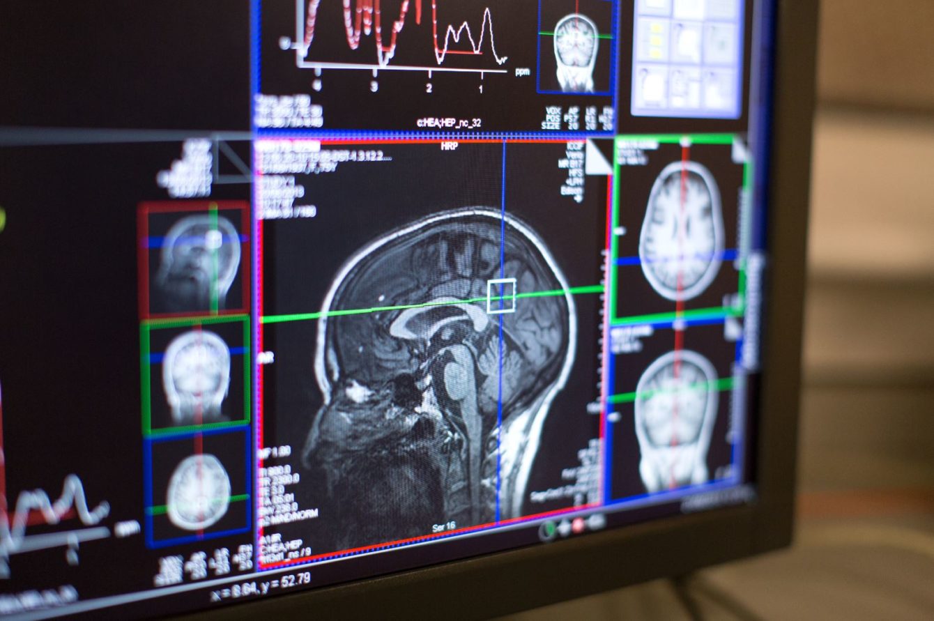
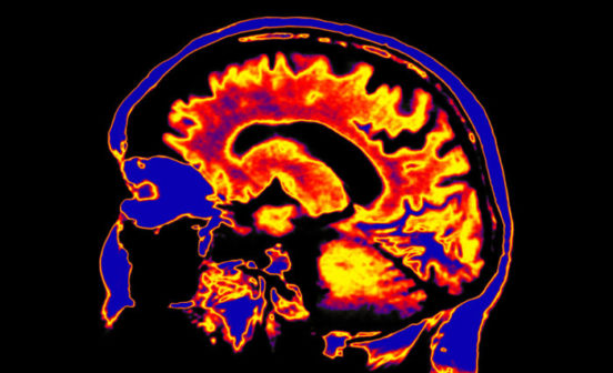
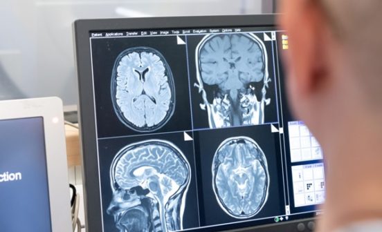
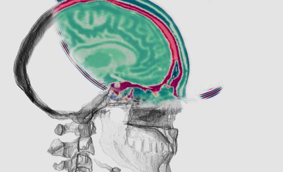
A team of researchers from Imperial College London, King’s College London, and the University of Oxford are developing new techniques which allow the brains of foetuses and babies to be scanned.
The Developing Human Connectome Project (dHCP) is a research project funded by a €15 million grant from the European Research Council, and is designed to map the growth of the baby’s brain during pregnancy and how it continues to progress after birth. The scans are carried out using Magnetic Resonance Imaging (MRI).
The aim of this research is to produce highly detailed information on brain development and to create a dynamic map of human brain connectivity linking together imaging, clinical, behavioural, and genetic information. The map of neural connections in the brain is called the “connectome” and scanning babies at a range of ages, from pregnancy until a few weeks after birth, will allow doctors and scientists to understand how the brain grows and also how problems may arise – this is the first research connectome study of early life. The project will help to understand how important diseases such as autism develop, or how problems in pregnancy affect brain growth.
Professor Daniel Rueckert is the dHCP Imperial College London Principal Investigator and part of the NIHR Imperial BRC Imaging Theme. “We have been developing novel approaches that help researchers by automatically analysing the rich and comprehensive MR images that are collected as part of dHCP” said Professor Rueckert.
The study has completed over 600 neonatal scans and has released a preliminary set of images which includes structural imaging, structural connectivity data, and functional connectivity data. Future data releases will consist of the image data with accompanying clinical, neurodevelopmental and genetic information.

