DiagnosticInnovationTherapeutic Improving results in dialysis patients through MRI scans
Dialysis is a procedure to remove waste products and excess fluid from the blood when the kidneys stop working properly. This is achieved by diverting blood to a machine to be cleaned and an arteriovenous fistula (AVF) is the surgically created connection that allows communication between an artery and vein for this treatment. However, AVF are susceptible to failure and ultimately stop working in approximately one in three patients leading to potentially life-threatening health problems.
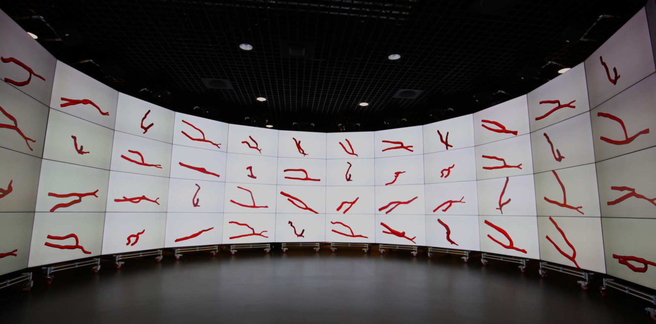
As part of this observational study, 60 dialysis patients that had the creation of an AVF (according to current surgical best practice in their wrist or upper arm) also participated in having a magnetic resonance imaging (MRI) scan on the day of surgery. The MRI is a type of scan that uses strong magnetic fields and radio waves to produce detailed images of the inside of the body. The aim of the MRI scans were to allow researchers to examine the three-dimensional geometry of AVF after surgery to measure the link between the structure of an AVF and how likely it is to fail.
The results showed that there was a significant variation in the three-dimensional structural conformation of AVF. Moreover, the researchers found that the more curved the AVF, the greater the success rates, and this also included AVF with larger angles between artery and vein. These results demonstrated that creating three-dimensional images of blood vessel structures prior to dialysis may predict how well a patient might take to treatment.

This study was the largest ever cohort of three-dimensional geometric reconstructions of AVF, and is a fantastic example of a collaboration between medics, aeronautical engineers and bioengineers from Imperial College London and Imperial College Healthcare NHS Trust using facilities supported by the BRC, including the Imperial College Clinical Imaging Facility.
Dr Richard Corbett, co-author from the Department of Medicine and Hammersmith Hospital, said: “Our findings show good reason to consider the 3D structure of AVFs when creating them in patients. We hope this will encourage surgeons who create these structures to think in three dimensions instead of two.”
The findings from this research will contribute towards preventing AVF failure in hospitals, improving health outcomes and saving money on operations for the NHS. The study team states that additional research is required to identify the best possible shape for the AVF in patients with kidney disease.
Read the original story by Caroline Brogan here.
Photo Credit: Peter Vincent


