DiagnosticInformatics ApproachesInnovationTherapeutic Radiomic classification method can help to improve treatment for lung cancer patients

Lung cancer is one of the most common and serious types of cancer, with a predicted 5-year survival rate of 8–13%. More than 85% of lung cancer cases are classified as non-small-cell lung cancers (NSCLC). A new study, led by researchers supported by the NIHR Imperial BRC, has discovered a machine learning methodology that predicts a 14-month survival difference for lung cancer patients undergoing chemo-radiotherapy.
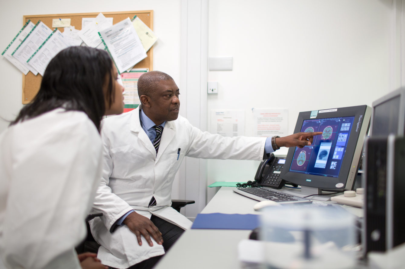
Imaging technology, such as positron emission tomography-computed tomography (also known as PET-CT) imaging, allows us to diagnose and determine the severity of or treat a variety of diseases, including cancer, heart disease, gastrointestinal, endocrine, neurological disorders and other abnormalities within the body.
For lung cancer, PET-CT images taken before treatment can also provide information to determine the stage of the disease (to understand the size, position and spread of the tumour) prior to treatment solutions such as surgery or chemo-radiotherapy. This is the primary method to determine treatment for NSCLC. However, this is an imprecise method and there is little information gathered on the expected development of the disease. If this type of detail can be uncovered, it will ultimately improve guiding treatment options available for patients.
Radiomics is the use of automated data analysis of large amounts of quantitative imaging features that are taken from medical images by using specific algorithms to uncover disease characteristics. This type of analysis can help to quantify a number of disease-specific variables and provide valuable information for personalised medicine.
A consortium of UK hospitals participated in this study and included Imperial College Healthcare NHS Trust, St James’s University Hospital in Leeds, Guy’s & St. Thomas’ Hospitals in London, the Royal Marsden Hospital in Sutton, Nottingham University Hospital, and Mount Vernon Hospital in Northwood. Data from the different hospitals were used to create imaging datasets for training and validation.
The training dataset included imaging data from 133 patients and was used to determine radiomic features. From 665 radiomic features identified, one combination of these features, labelled as FVX (feature vector), was selected and tested on the validation dataset, which comprised of scans from 204 patients. Results demonstrated that FVX predicted a 14-month survival difference in the validation group without knowing the stage of the cancer. This means that quantifying the variation within lung tumours – using PET radiomics – can provide decision support for tailoring therapies to the patient.
Professor Eric Aboagye, NIHR Imperial BRC Imaging Theme Lead said “the landscape of lung cancer treatment has changed over the last few years with the advent of immunotherapy and other molecularly targeted therapies. The ability to group patients according to the condition and likely development of their medical condition allows clinicians to select the best therapies and/or intensity of treatment.” He added, “For the basic and computational biologists this type of work provides further substrate for more research, to address the question why two patients with the same stage-disease have remarkably different outcomes?”
This multi-institutional study demonstrates valuable results highlighting the opportunity to re-purpose imaging data collected in various NHS hospitals through routine scans to obtain predictive information to guide treatment options.


