AwardInnovationTherapeutic Navigation with enhanced surgical vision for robot-assisted operations
Brain tumours kill more children and adults under the age of 40 than any other cancer. This is because surgeons do not have yet the tools to help them navigate through the delicate paths of the brain, accurately and safely. Being able to clearly identify cancerous tissue during surgery is an important step towards improving outcomes and quality of life for people undergoing brain surgery.
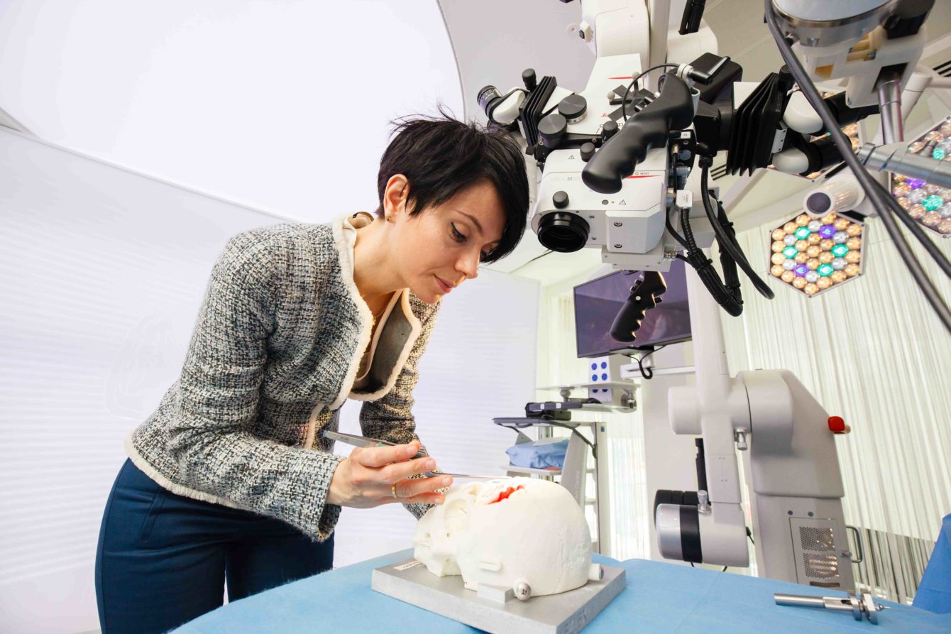
Dr Stamatia (Matina) Giannarou is a Royal Society University Research Fellow and a Lecturer in Surgical Cancer Technology and Imaging at the Hamlyn Centre for Robotic Surgery, and a researcher within the Surgery & Surgical Technology BRC Theme. Her research focuses on enhanced surgical vision for intraoperative navigation in minimally invasive and robot-assisted operations. Recently, she won “The President’s Award for Outstanding Early Career Researcher 2017” at Imperial College London. She has received several best paper awards and has been invited to present her work at international workshops and symposia and has been selected as a member of the IdeasLab of Imperial College London on the “Frontiers of Imaging” at the World Economic Forum Annual Meeting of the New Champions 2016 in Tianjin, China. We caught up with her to find our more about her work and career.
How did you become interested in your field of work?
Throughout my early career development, my main research interest has been in surgical robot vision. As part of my fellowship, I decided to focus my research on developing surgical navigation tools to improve the efficacy and safety of tumour resection in neurosurgery. This is because after discussing extensively with neurosurgeons and observing live neurosurgical operations, the need for diagnostic and therapeutic tools that can improve the safety and accuracy of neurosurgical oncology became apparent. Furthermore, the brain is the most powerful organ in the human body and when something is going wrong with the brain, it can be a personal tragedy because it changes who we are, what we think, what we feel.

What are the major current challenges in management/treatment of brain tumours?
Surgery on tumours residing within the brain is particularly demanding, and the prognosis for patients afflicted with such tumours remains very poor. For example, glioblastoma multiforme (GBM), the most frequent primary malignant brain tumour, has a median survival of less than 18 months despite multi-modal therapy with surgery, radiotherapy and chemotherapy. Intrinsic brain tumours are highly infiltrative making it difficult to distinguish tumour tissue from surrounding tissue. Moreover, it is imperative to preserve unaffected brain tissue, which is delicate, often eloquent, and has little capacity for regeneration. The corollary is that technologies that allow for improved identification of cancer tissue, and more precise and delicate resection, may improve patient outcomes. Current state-of-the-art technologies used to facilitate brain tumour identification such as neuronavigation, iMRI and fluorescence imaging have significant limitations intraoperatively or can not provide complete tumour identification. Therefore, the need for hybrid diagnostic and therapeutic tools that can improve the safety and accuracy of oncological surgery becomes apparent.
Could you tell us about your research?
The aim of my research is to integrate multimodal intraoperative imaging and navigation technologies into a cognitive robotic platform to allow for more accurate tumour margin delineation. This will enable accurate and highly personalised in vivo tissue characterisation with the aim of improving both the efficacy and safety of tumour resection. My work focuses on the intraoperative surgical navigation, the anatomy-specific robotic tissue scanning with imaging probes and the tissue characterisation with on-line diagnosis support. These research objectives are in response to the current paradigm shift and clinical demand in bringing cellular and molecular imaging modalities to an in vivo and in situ setting to allow for real-time tissue characterisation and surgical guidance. A key application of this cognitive robotic platform is the resection of glioblastoma multiforme but its versatile nature makes it suitable for any cancer resection procedure. The development of the above platform will have a significant impact on the surgeon’s sensing, completeness and safety during tumour resection and ultimately on the patient’s quality of life.
What are you proudest of in your research or career so far?
I am proud for having been awarded the Royal Society University Research Fellowship which has allowed me to work on an interesting research area and to develop my research independence. Through this project, I have built research collaborations with clinical and engineering teams which have led to the submission of collaborative papers and research grant proposals.
What is the biggest challenge you faced in your career?
The biggest challenge I face in my academic career is to find the right balance between spending quality time with my family and working towards fulfilling my career goals. To achieve this, I am trying to allocate several hours every week to my family and friends as, I do feel that it boosts my performance at work and helps me redirect my mind away from work.
What are you most excited about as a researcher?
My work as a researcher focuses on an exciting area not only because it enables me to tackle challenging research objectives but mainly because it has the potential to be translated to clinical practice and bring significant impact to people and the society. In addition, working on an interdisciplinary research area, I enjoy collaborating both nationally and internationally with engineers, scientists, clinicians and medical researchers.


