PreventionTherapeutic New insights into heart muscles could provide explanations for heart diseases

Researchers at the MRC Laboratory of Medical Sciences (LMS) and Imperial College London have combined imaging and genetic data from almost 50,000 people to reveal how our genes influence a network of heart muscle fibres called cardiac trabeculae. While these muscles play a key role in healthy heart activity, increased trabeculation is a feature of heart disease. The findings provide new insights into the heart’s adaptive response to disease.
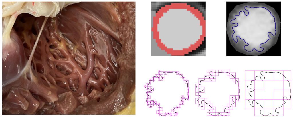
Cardiac trabeculae are a network of muscle fibres found in our circulatory system, lying between fast-flowing blood inside the heart and the contracting heart muscle. As long ago as the 16th Century, the artist Leonardo Da Vinci was the first to sketch these muscles, speculating at the time that they warm the blood as it flows through the heart. Yet it is only now that we are beginning to truly understand how important they are to human health.
The network of fibres is thought to play a role in the efficient pumping and electrical activity of the heart, and they adapt to changes in demand placed on the heart. Increased trabeculation can be seen in healthy people, including pregnant people and those who do extreme sports. The trabeculation pattern can return to normal after the demand on the heart stops.
Importantly, increased trabeculation is also a feature of diseases, such as cardiomyopathy – heart muscle diseases that are the leading cause of heart transplants and sudden death. The presence of trabeculation on scans has created controversy amongst doctors for many years, leading them to question whether disease causes trabeculation or trabeculation causes disease. The research, published in Nature Cardiovascular Research and led by Dr Kathryn McGurk working with Professor Declan O’Regan, Head of the Computational Cardiac Imaging group at the LMS and a key member of the NIHR Imperial BRC Cardiovascular Theme, begins to answer this clinical question.
The interdisciplinary team used sophisticated machine learning to measure and analyse trabeculation on cardiac MRI images from almost 50,000 people’s hearts from UK Biobank – a large-scale biomedical database and research resource. By comparing the MRI imaging data with genetic data for each participant, the team identified a large repertoire of genes responsible for the development and function of trabeculae. Many of these genes overlap with genes that cause cardiomyopathy. The team determined that interactions between both rare and common genetic variants determine how trabeculae in the heart look and how diseases may evolve.
Taken together, the findings suggest that understanding the genetics of trabeculation adds a missing piece of the puzzle in the drivers of variability in cardiomyopathy and what contributes to the diversity in the appearance of trabeculation in the heart.
Professor O’Regan said, “Trabeculae lie in a critical part of the heart and we discovered a repertoire of developmental genes that are associated with their appearance. These genes may also play an important role in explaining the diverse manifestations of inherited heart diseases”
Dr McGurk added, “We’re beginning to understand the genetic, clinical, lifestyle and other factors that influence these structures. They can occur early in the pathogenesis of heart disease and progress with disease, making them useful markers of cardiac remodelling when identified in individuals at particular risk of heart disease.”


